 Dr. Xiangrui “Taylor” Zeng
 2023 Human Biology Project Fellow
Written by: Dr. Tam Maiuri
Edited by: Dr. Kaitlyn Deschamps
Seeing Beneath the Surface: How Artificial Intelligence Could Reveal Hidden Clues in the HD Brain
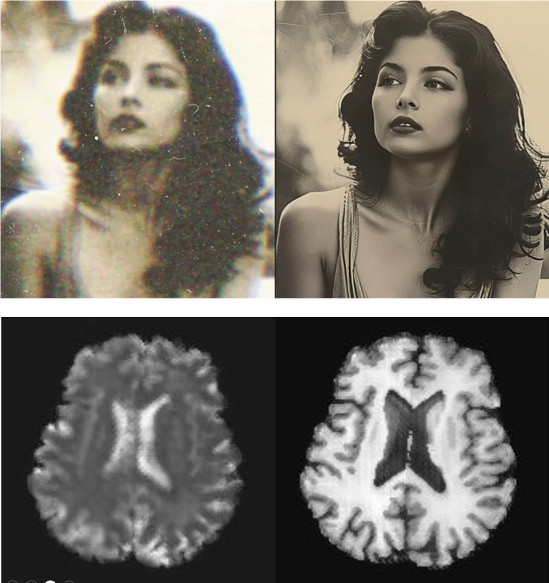
Most brain scans using MRI look like a blur of grey and white, making it difficult to see the details. But Dr. Xiangrui (“Taylor”) Zeng, a postdoctoral researcher at Massachusetts General Hospital and Harvard Medical School, is teaching computers to sharpen that picture, revealing fine layers of the brain that may hold key insights into how Huntington’s disease (HD) begins and progresses.
Taylor’s work is supported through the HDSA Human Biology Project, a research program that funds studies focused directly on HD in people. His project, Integrating clinical MRI and ultra-high-resolution ex vivo MRI to model laminar anomaly progression in Huntington’s disease, uses artificial intelligence to map which layers of the brain’s cortex are most vulnerable to HD and at what stage they become affected.
From Curiosity to Collaboration
Taylor’s interest in HD began to grow from a broader fascination with how the brain develops and deteriorates. “I was deeply interested in neurodevelopmental and neurodegenerative diseases since college,” he explained. “I’m curious about how neurons develop and how they deteriorate, and how that affects cognition and behaviour.”
He first explored psychiatric conditions like schizophrenia and depression during his neuroscience studies, and then earned a PhD in computational biology at Carnegie Mellon University. Taylor became increasingly interested in diseases that connect small, molecular changes to real, measurable changes in brain structure.
This curiosity led him to the HD field—a space defined not only by scientific challenges but by a unique sense of community. “Alzheimer’s disease is widely studied, but Huntington’s is rarer,” he said. Because of this, every HD dataset is especially valuable. Taylor explained that researchers today are fortunate to have access to shared brain scans, donated tissue, and collaborative studies built through years of dedicated partnership from the HD community.
Now at Mass General, Taylor is collaborating with Dr. Diana Rosas, a longtime HDSA partner whose work has shown that the brain’s cortex begins thinning even before HD symptoms appear. Her research, supported by HD families through participation in clinical studies and brain donation, laid the foundation for Taylor’s project. As he put it, this field “offers very important biological insights into the disease—but we need ways to apply that knowledge in living patients.”
Turning Blurry Brain Scans into Layer-by-Layer Maps
The thin outer shell of the brain, called the cortex, is made up of multiple layers, each packed with specialized neurons. In HD, those neurons begin to die, leading to cortical thinning. But this loss doesn’t happen evenly. Some layers shrink first, while others remain intact for years. Understanding which layers go first could reveal how HD progresses throughout the brain.
The challenge is that traditional clinical MRI scans can’t show these fine structural details. To tackle this problem, Taylor is using AI trained on ultra-high-resolution images of donated HD brains. These scans capture details that can’t be measured in living patients. By teaching AI to recognize the subtle differences between cortical layers in these high-resolution images, Taylor can then apply the same pattern recognition to standard hospital MRIs, effectively “boosting” their resolution.
He compared it to enhancing an old photo: “Fifty years ago, we had low-resolution grey-scale images; now we can recolor and enhance them. We’re doing something similar with the cortex, but in 3D.”
Science That Builds on Shared Tools and Shared Generosity
Taylor’s work stands on decades of collaboration. His mentor, Dr. Bruce Fischl, developed FreeSurfer, the openly accessible software used by tens of thousands of neuroscientists worldwide to analyze brain MRI data. FreeSurfer makes it possible to measure the thickness and shape of different cortical regions—precisely the kind of data Taylor’s AI models depend on.
It’s a perfect example of how progress in the HD field builds: each project benefits from the tools, data, and generosity of those who came before it.
This generosity includes the profound contribution of brain tissue donation. The high-resolution scans Taylor uses as “training data” for his AI program exist because of people who chose to donate their brains to science. And their contributions don’t expire. Because AI is advancing so quickly, data donated today can be re-examined year after year, with newer algorithms uncovering information that simply couldn’t be detected before.
A Sharper Picture of the Future
Taylor’s goal is to build a model that shows how cortical layers change over time in HD, from the earliest, pre-manifest, to the late stage. In the next five years, he plans to continue this work in both HD and Alzheimer’s disease, using insights from each to strengthen the other. Both diseases involve subtle, layer-specific changes in the cortex long before symptoms appear, so advances in one field can strengthen the tools used in the other.
If successful, this work could yield a new kind of imaging biomarker—one sensitive enough to detect the earliest signs of HD-related brain change and to measure whether treatments are truly protecting neurons.
But Taylor also believes that advancing HD research takes more than technology. “Almost everyone knows Alzheimer’s or Parkinson’s, but few have even heard of Huntington’s,” he said. The disease’s rarity keeps it invisible to the broader public, which limits the resources directed toward it. He hopes that greater awareness will help ensure that HD receives the same scientific and public investment that more popular brain diseases do.
Science in Practice
Outside the lab, Taylor practices daily meditation. It’s a habit that reflects his belief in maintaining brain health—a topic his mentor, Bruce Fischl, has studied extensively. Fischl’s research showed that long-term meditation is linked to increased cortical thickness, the very feature Taylor now measures in his imaging work. It’s a small, personal way of putting the science he studies into practice.
Fast Facts
HDSA Program: Human Biology Project
Institution: Massachusetts General Hospital / Harvard Medical School
Mentor: Dr. Bruce Fischl
Collaborator: Dr. Diana Rosas
Focus: AI-based modeling of cortical layer changes in HD
Why it matters: Could provide an earlier, more sensitive imaging biomarker for HD progression
Fun fact: His algorithms will be integrated into FreeSurfer, a neuroimaging platform used by over 60,000 researchers worldwide
Dr Zeng’s project is a reminder of what happens when technology, collaboration, and community generosity come together: a sharper view of the HD brain—and of the future of HD research.